What is Intracranial Hemorrhage?

Intracranial Hemorrhage (ICH) is a fatal hemorrhagic stroke condition characterized by bleeding within the skull. It represents a critical medical emergency that requires rapid diagnosis and immediate intervention to prevent permanent neurological damage or death.

ICH can occur in various locations within and around the brain, each presenting unique clinical challenges and requiring specific treatment approaches. The prognosis and treatment strategy depend heavily on the location, size, and cause of the hemorrhage.

Anatomical Context: The Meninges

Understanding ICH subtypes requires knowledge of the brain's protective layers, called the meninges. From outermost to innermost:

  • Skull (Calvarium): The hard bone protecting the brain
  • Dura Mater: Tough, outermost meningeal layer attached to the skull
  • Arachnoid Mater: Middle layer forming a web-like structure
  • Subarachnoid Space: Space containing cerebrospinal fluid and blood vessels
  • Pia Mater: Thin innermost layer adhering to brain surface
  • Brain Parenchyma: The brain tissue itself
  • Ventricular System: Interconnected cavities containing cerebrospinal fluid
Meninges Anatomy Illustration

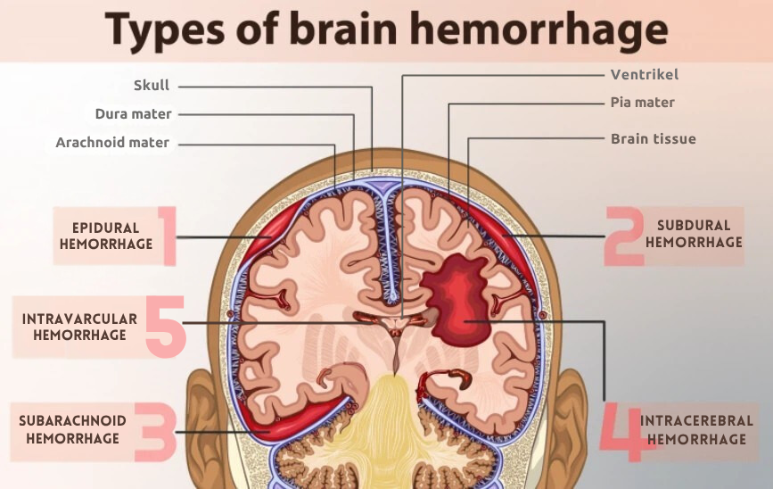
Diagram showing layers from skull to brain tissue with ICH locations

Five Detected ICH Subtypes

This system detects five distinct types of intracranial hemorrhage, each with unique characteristics, causes, and clinical implications.

Epidural Hemorrhage (EDH)

Traumatic

Bleeding between the skull (calvarium) and the dura mater, typically caused by arterial injury following skull fracture.

Common Causes:

  • Skull fracture with middle meningeal artery tear
  • Severe head trauma or impact
  • Motor vehicle accidents
  • Falls from height

Clinical Significance:

Often presents with the classic "lucid interval" - brief unconsciousness, followed by apparent recovery, then rapid deterioration. Requires urgent neurosurgical intervention. Excellent prognosis if treated before herniation.

Subdural Hemorrhage (SDH)

Common

Blood collection between the dura mater and arachnoid mater, commonly caused by tearing of bridging veins.

Common Causes:

  • Head trauma (even minor in elderly)
  • Falls, especially in elderly patients
  • Anticoagulation therapy
  • Brain atrophy creating space for blood accumulation
  • Chronic alcohol abuse

Clinical Significance:

Can be acute (symptoms within 72 hours) or chronic (develops over weeks). Particularly common in elderly population. May require surgical drainage depending on size and symptoms.

Subarachnoid Hemorrhage (SAH)

Critical

Bleeding in the subarachnoid space between the arachnoid and pia mater, most commonly caused by ruptured cerebral aneurysm.

Common Causes:

  • Ruptured cerebral aneurysm (80-85% of cases)
  • Arteriovenous malformation (AVM)
  • Head trauma
  • Bleeding disorders

Clinical Significance:

Classic presentation: sudden "thunderclap" headache - worst headache of one's life. High mortality rate (40-50%). Risk of rebleeding and delayed vasospasm. Requires immediate neurosurgical evaluation and aneurysm securing.

Intraventricular Hemorrhage (IVH)

Serious

Bleeding within the brain's ventricular system where cerebrospinal fluid is produced and circulates.

Common Causes:

  • Extension from intraparenchymal hemorrhage (most common)
  • Primary intraventricular bleeding (rare)
  • Arteriovenous malformation rupture
  • Aneurysm rupture

Clinical Significance:

Can obstruct CSF flow leading to acute hydrocephalus. High mortality rate (50-80%). May require urgent ventricular drainage. Presence of IVH significantly worsens prognosis of ICH.

Intraparenchymal Hemorrhage (IPH)

Most Common

Bleeding directly within the brain tissue (parenchyma), representing the most common form of spontaneous intracerebral hemorrhage.

Common Causes:

  • Chronic hypertension (most common, 50-60% of cases)
  • Cerebral amyloid angiopathy
  • Anticoagulant therapy complications
  • Head trauma
  • Brain tumors

Clinical Significance:

Accounts for 10-15% of all strokes with 30-day mortality of 35-52%. Presents with sudden focal neurological deficits depending on location. Treatment depends on size, location, and patient's clinical condition.

Why Early Detection Matters

⏱️

Time-Critical Condition

ICH progression can be rapid. Early detection within the "golden hour" significantly improves survival rates and neurological outcomes.

🏥

Treatment Planning

Accurate subtype identification guides appropriate treatment strategy - surgical intervention, medical management, or observation.

📊

Prognosis Assessment

Different ICH subtypes have varying mortality rates and recovery trajectories. Early classification helps in patient counseling and resource allocation.

⚠️ Educational Purpose Only

This page provides educational information about intracranial hemorrhage for academic and learning purposes. It is not intended to provide medical advice or replace consultation with qualified healthcare professionals. All medical decisions should be made by licensed physicians based on comprehensive patient evaluation and clinical judgment.